Imagining a better way through advanced imaging
By GuideWell Emergency Doctors
Share:
June 17, 2024
It’s not hard to imagine why doctors and medical professionals value cutting-edge imaging tools. Thanks to detailed images from advanced tests (like the CT scan) doctors can quickly and accurately rule out or rule in everything from bone fractures and internal injury to life-threatening conditions (like cancer, heart disease, and so on). At GuideWell Emergency Doctors, imaging is everything.
GuideWell Emergency Doctors is well equipped with the latest imaging technology to treat patients (by walk-in or appointment) with the highest level of care. Rest assured, we have the same kind of advanced on-site imaging services you’d find at a hospital emergency room (ER), including:
You can also count on us for the medical expertise needed to interpret detailed images accurately. In fact, all of our physicians (Board-Certified Emergency Medicine Doctors) have had at least five years of ER experience before joining GuideWell Emergency Doctors.
Likewise, our certified technicians for both X-ray and CT scans (working alongside U.S.-based, board-certified radiologists) provide final CT scan reads in 30 minutes or less and final X-ray and ultrasound reads in 60 minutes or less.
At GuideWell Emergency Doctors, we’re committed to providing the same high-level emergency care as a hospital ER but in 1/2 the time1 at 1/3 the cost2 of an average ER visit.
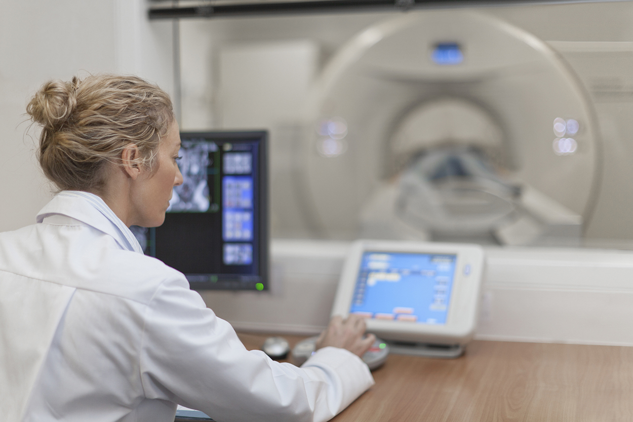
Technically known as computed tomography, a CT scan is a diagnostic imaging procedure that combines X-ray and computer technology to produce precise, detailed images of the body.
During a CT scan, you lie inside a donut-shaped machine while a scanner spins around your body to capture precise, detailed images (from cross-sectional points of view). Each rotation is known as a slice. Each slice contains multiple detailed images. During a CT scan, a radiologist can look at the detailed images individually or use a computer to generate 3D images for analysis.
Never had a CT scan? Take comfort in the fact that it’s a quick and painless medical procedure. Not only is a CT scan fast (much faster than an average magnetic resonance imaging or MRI scan), but it’s also considered a safe diagnostic tool, given that a CT scan involves tiny amounts of radiation — about the same amount you’d otherwise be exposed to over a three- to five-year period.3
CT scans can provide detailed images of everything from bones and blood vessels to organs, muscle, and soft tissues. A doctor might order a CT scan for numerous reasons (such as confirming a bone break or identifying the source of internal pain). Of course, not every medical situation requires a CT scan. A doctor might well find that an X-ray is all they need to rule in or out bone fractures, for example. You might be wondering…
X-ray technology is the most commonly used diagnostic imaging test. Even if a doctor suspects you’ll need more detailed images (through a CT or MRI scan), there’s a good chance, you’ll get an X-ray first. Technically put, X-rays use invisible electromagnetic energy beams to produce images of internal tissues, bones, and organs (either on film or digitally). X-rays are a form of radiation. When electromagnetic beam pass through the body, bone and other densities block the radiation and look white on the X-ray (compared with less dense tissues that appear gray).
| X-ray | CT scan |
|---|---|
| 2D imaging | 3D imaging |
| Mainly for viewing bones, cancer, pneumonia | Mainly to see/diagnose organ and tissue conditions |
| Uses radiation to produce images | More powerful than X-ray; little radiation involved |
| Common/widely used | 360-degree view of body |
Wonderful tools in advanced 3D imaging, CT scan and MRI scan technology both capture detailed images within your body.
Unlike a CT scan, which uses X-ray technology, an MRI scan uses radio waves and a powerful magnet to create detailed images.
Depending on a patient’s unique situation, a doctor may opt for a CT scan or an MRI scan. For example, a CT scan is highly effective for diagnosing stroke, blood clots, and internal injury (including fracture), while an MRI scan may be the best choice for examining damage to soft tissues or assessing the presence of certain diseases.
| CT scan | MRI scan |
|---|---|
| 3D detailed images | 3D detailed mages |
| Uses computed tomography (X-ray and computer) | Uses radio waves and powerful magnet |
| Faster results (readily used in ERs) | Longer procedure/wait times |
|
Often used to diagnose:
|
Often used to diagnose:
|
Just like you’d find in a hospital ER, GuideWell Emergency Doctors is equipped with the latest in CT scan technology. If our Board-Certified Emergency Medicine Doctors feel that an MRI scan would be the best course of action for you, our caring team will be happy to help you get set up for an MRI scan at an imaging center near you.
In addition to CT scans and X-ray imaging, GuideWell Emergency Doctors also offers ultrasound imaging services.
At GuideWell Emergency Doctors, we also use ultrasound (or sonography/ ultrasonography) to care for our patients. Ultrasound is a noninvasive imaging test that uses high-frequency sound waves to take detailed images (picture and video) of internal organs or other soft tissues. Unlike with X-ray, there’s no radiation involved.
Most ultrasound tests are done using a device (transducer) on your skin. Some ultrasound procedures (like pelvic ultrasound) require placing a small device inside your body.
Pregnancy (prenatal) ultrasound
A widely used form of imaging used to:
Diagnostic ultrasound
There are a wide range of diagnostic ultrasounds to help doctors diagnose possible internal issues. They include:
Guidance ultrasound
Ultrasound can also be vital in helping doctors precisely and safely perform procedures, including those that require guiding a needle. For example:
If it’s advanced imaging you need, you’re in capable hands at GuideWell Emergency Doctors.
Though not an imaging procedure, per se (since it does not take detailed images), EKGs (also known as electrocardiograms) are a vital on-site diagnostic test we offer at GuideWell Emergency Doctors. EKG is also one of the easiest, most effective tests used to evaluate the heart.7
During an EKG, electrodes (small, plastic patches stuck to the skin) are placed on the chest, arms, and legs. A patient’s heart activity is transmitted from those electrodes (through lead wires) to an EKG machine that monitors, measures, and prints out results.
A doctor may order an EKG if a patient is experiencing:
EKGs can truly be a lifesaver by quickly diagnosing:
No one likes to think about having a medical emergency, but isn’t it comforting to know that when and if you do, you have an option that can save you time and money compared to a hospital ER? Providing ER-level advanced imaging services is one more way your nearby GuideWell Emergency Doctors is leading the way in emergency and urgent care.
(sources)
1CMS.gov data measuring timely and effective emergency department care by state compared to GuideWell Emergency Doctors’ visit data from 2023 showing time from check-in to discharge is 80 minutes on average.
2Cost is based on health plan actuarial analysis performed in 2021 that compared cost of hospital-based ER visit versus a GuideWell Emergency Doctors visit
3https://americanhealthimaging.com/mri-vs-ct-scans
4https://www.envrad.com/difference-between-x-ray-ct-scan-and-mri/
5https://practiceplusgroup.com/knowledge-hub/ct-scan-vs-mri-difference/
6https://my.clevelandclinic.org/health/diagnostics/4995-ultrasound
7https://www.hopkinsmedicine.org/health/treatment-tests-and-therapies/electrocardiogram